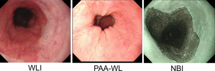
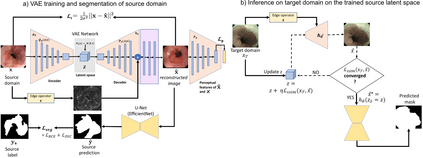
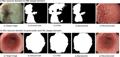
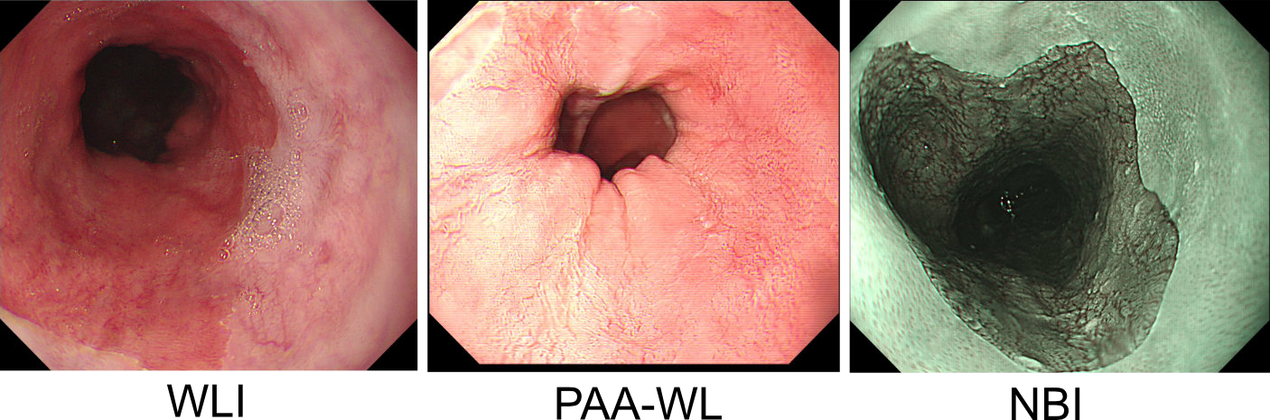
Barrett's oesophagus (BE) is one of the early indicators of esophageal cancer. Patients with BE are monitored and undergo ablation therapies to minimise the risk, thereby making it eminent to identify the BE area precisely. Automated segmentation can help clinical endoscopists to assess and treat BE area more accurately. Endoscopy imaging of BE can include multiple modalities in addition to the conventional white light (WL) modality. Supervised models require large amount of manual annotations incorporating all data variability in the training data. However, it becomes cumbersome, tedious and labour intensive work to generate manual annotations, and additionally modality specific expertise is required. In this work, we aim to alleviate this problem by applying an unsupervised domain adaptation technique (UDA). Here, UDA is trained on white light endoscopy images as source domain and are well-adapted to generalise to produce segmentation on different imaging modalities as target domain, namely narrow band imaging and post acetic-acid WL imaging. Our dataset consists of a total of 871 images consisting of both source and target domains. Our results show that the UDA-based approach outperforms traditional supervised U-Net segmentation by nearly 10% on both Dice similarity coefficient and intersection-over-union.
翻译:巴雷特的眼科成像(BE)是食道癌的早期指标之一。BE病人受到监测并接受消化疗法,以尽量减少风险,从而使它能够精确地识别BE区域。自动分解可以帮助临床内分解科医生更准确地评估和治疗BE区域。BE的内窥镜成像除了常规白光(WL)外,还可以包括多种模式。监督模型需要大量人工说明,将培训数据的所有变异性纳入培训数据中。然而,它变得烦琐、烦琐和劳动密集型工作,以生成手动说明,并需要更多特定模式的专门知识。在这项工作中,我们的目标是通过应用不受监督的域适应技术(UDA)来缓解这一问题。在这里,UDA接受关于作为源域的白光内镜学图像的培训,并且非常适合将不同成像形式作为目标域,即窄带成色成像和后电酸性酸成像成像。我们的数据集由总共871张图像组成,包括源和目标域和目标域。我们的目标是通过一种不受监督的域适应的域适应的UDA分位法,以近的UDA为U-BI-DA的分位。